RETINAL SURGERY

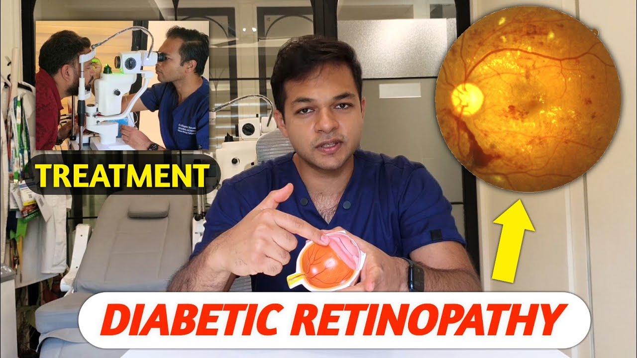
Diabetic Retinopathy

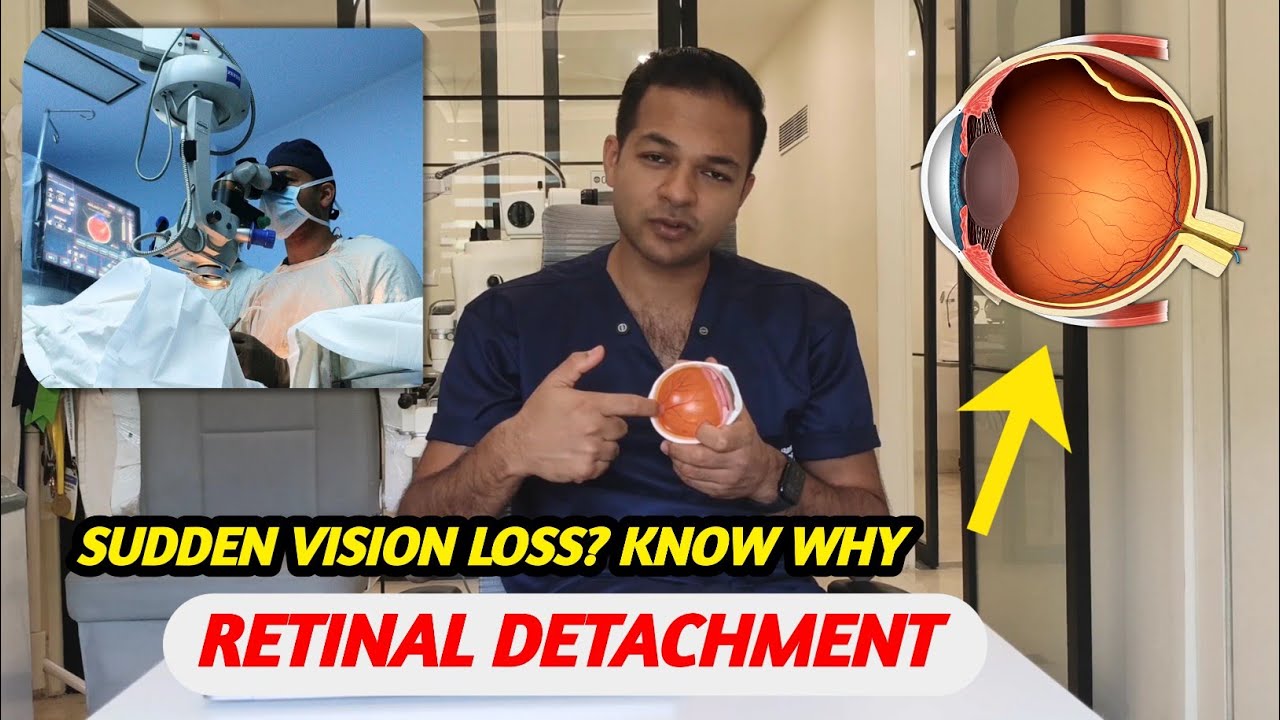
Retinal Detachment

Age-Related Macular Degeneration (AMD)

Macular Hole

Retina Surgery in Delhi — Retinal Detachment, Vitrectomy & Macular Hole Treatment
Expert Retina Care with Dr. Mayank Bansal, Retina Specialist
Many patients arrive at the clinic feeling anxious after hearing the term "retinal detachment," and that's understandable, it sounds alarming. So it helps to start with the basics: the retina is the thin layer of light-sensing tissue lining the back of the eye. It's what actually captures the image and sends it to the brain. When it tears, detaches, or gets damaged in some other way, vision can change quickly. The encouraging part is that retinal problems are also among the most treatable conditions in ophthalmology, provided they're caught and acted on in time with the right retina surgery.
Dr. Mayank Bansal is a retina specialist who has built his practice specifically around retina surgery, both diagnosis and surgical treatment. His approach centers on making sure patients actually understand what's happening in their eye, not just receiving a treatment plan without context. This page is meant to offer that same grounding: what retina surgery involves, how the different retinal detachment and vitrectomy procedures work, and what recovery really looks like.
What Is Retina Surgery?
"Retina surgery" isn't a single operation, it's a family of procedures, each suited to a different kind of retinal problem, from retinal detachment repair to vitrectomy surgery and macular hole treatment. Every one of them, though, works toward the same outcome: reposition and repair the retina, and preserve as much vision as possible.
Here's something that often surprises patients, retinal tissue doesn't heal on its own the way skin does. Once it tears or detaches, it stays that way until it's surgically corrected. This is why timing matters so much when it comes to retinal detachment surgery. Clinical research on macula-involving detachments has found that patients treated within the first few days of losing central vision tend to have better visual outcomes than those treated after a longer delay, which is a big part of why most detachments are treated as same-week, if not same-day, priorities.
Retinal Conditions Commonly Treated with Retina Surgery
Types of Retina Surgery: The Surgical Options
There isn't one "right" way to fix a retinal problem. The choice of retina surgery procedure depends on the type of break or damage involved, its location, whether the vitreous is pulling on it, and the overall health of the eye.
Pars Plana Vitrectomy (PPV)

This is the most frequently used retina surgery procedure, and for good reason: it suits a wide range of situations, including breaks located toward the back of the eye, multiple breaks in different areas, giant retinal tears, dense vitreous hemorrhage obscuring the view, and detachments complicated by scar tissue (proliferative vitreoretinopathy). Working through a few incisions smaller than the tip of a pen, the surgeon removes the vitreous gel that's pulling on or clouding the retina, then repairs the retina directly from inside the eye.
At the end of the vitrectomy surgery, the eye is typically filled with a gas bubble, or, for more complex repairs, silicone oil, to hold the retina flat while it heals.
Scleral Buckle Surgery

Instead of working inside the eye, this retinal detachment surgery technique places a thin, flexible silicone band around the outside of it. The band gently indents the eye wall, relieving the traction that pulled the retina away in the first place, and is usually paired with laser or freezing treatment (cryotherapy) to permanently seal any tears. Among well-selected cases, a single scleral buckle procedure achieves successful reattachment in roughly 80–90% of eyes. The band sits beneath the surface tissue of the eye, it isn't visible from outside, and it generally remains in place permanently.
This remains an excellent retina surgery option for certain detachments, particularly less complex ones and in younger patients with a clear vitreous.
Pneumatic Retinopexy

For carefully selected, less complex retinal detachments, this can be performed in the clinic rather than the operating room. A small bubble of gas is injected into the eye, and with specific head positioning over the following days, the bubble presses the detached retina back against the eye wall while laser or cryotherapy seals the tear.
Laser Photocoagulation

Used more broadly for sealing retinal tears or as part of diabetic retinopathy treatment for abnormal, leaking blood vessels, this creates a ring of small, controlled burns that scar down and effectively weld the retina in place.
Getting Technical - On Instrument Gauge: 23G vs 25G vs 27G

One detail patients rarely ask about, but that actually matters quite a bit, is the size of the instruments I'm working with, measured in "gauge." The three sizes I choose between each have a different feel in surgery:
-
23G is the sturdiest of the three. It gives solid, confident control when working on something delicate, which matters in tougher cases. The trade-off is that the incision is slightly bigger, so entry can be a touch more traumatic to the eye, and occasionally need to place a suture to close it properly.
-
25G is my usual middle ground. There's still enough rigidity to control the instruments precisely, but the incisions are small enough that they typically seal themselves by the end of surgery, no stitches required.
-
27G is the finest gauge available, and the incisions from it almost always close on their own. But there's a real trade-off: the instruments are noticeably more flexible, they can bend under resistance inside the eye, which honestly makes certain maneuvers harder to execute cleanly, even for an experienced surgeon.
Depending on the underlying retinal disease, additional steps may include:
-
Removal of vitreous hemorrhage (bleeding inside the eye)
-
Delicate peeling of scar tissue and tractional membranes
-
Internal limiting membrane (ILM) peeling
-
Epiretinal membrane (ERM) peeling
-
Repair of retinal tears
-
Reattachment of a detached retina
-
Endo laser photocoagulation
-
Injection of specialized intraocular gases or silicone oil to support retinal healing
Every operation is customized to your diagnosis, retinal anatomy, and visual requirements. There's no one-size-fits-all retina surgery.
Barrage Laser Retinopexy

This retina surgery approach is used somewhat differently, for a localized, subclinical retinal detachment or an isolated tear that hasn't yet progressed to a full detachment. A ring of laser burns is placed around the area to "wall off" the problem and prevent it from spreading further. It's usually done under topical anesthesia in the clinic, though patients need to understand it doesn't guarantee the area won't still progress and require further treatment later.
Cryotherapy

A freezing probe applied to the outside of the eye creates a similar sealing effect to laser, often used in combination with a scleral buckle or pneumatic retinopexy.
Retina Surgery Procedure: What Surgery Day Involves
Most retina surgery procedures are performed under local anesthesia with sedation, so the patient is comfortable and pain-free without needing general anesthesia. Depending on complexity, retina surgery typically takes 30 minutes to two hours, and the large majority of patients go home the same day.
Before any of that, Dr. Bansal carries out a detailed retinal examination, frequently supported by OCT (optical coherence tomography) imaging or ultrasound, to map precisely what's happening in the eye and plan the most appropriate surgical approach.
Retina Surgery Recovery Time & Aftercare
The first few days after retina surgery typically bring mild soreness, redness, and blurred vision, this is normal. Patients usually wear a protective eye shield, especially while sleeping, to avoid accidental pressure on the eye.
If a gas bubble was placed, a specific head position, often face-down, may be needed for several days, so the bubble stays in contact with the repair site. It's not the most comfortable part of recovery, but it plays a real role in how well the repair holds.
Vision often looks worse before it improves. This catches people off guard, they expect clarity right away and instead see a shifting, hazy bubble. That's expected. As the eye heals and the gas gradually absorbs over several weeks, vision typically continues to improve, often over a period of weeks to a few months.
Activity is restricted for a while. Heavy lifting, strenuous exercise, and swimming are usually paused for a few weeks. If a gas bubble is present, air travel and high-altitude locations must be avoided entirely until it has fully absorbed, since pressure changes at altitude can be genuinely dangerous with a gas-filled eye.
Follow-up visits are not optional extras. They're how early signs of re-detachment, elevated eye pressure, or other complications get caught before they become bigger problems.
Most people resume light daily activities within a week after retina surgery. Full visual recovery generally takes four to eight weeks, sometimes longer for more complex repairs.
Why Choose Dr. Mayank Bansal as Your Retina Surgeon
-
Years of focused experience in medical and surgical retina care, not general ophthalmology with retina as a secondary interest
-
Access to detailed diagnostic imaging that maps the retina precisely before surgery is ever discussed
-
A surgical plan, and instrument choice, matched to each patient's specific case rather than a standard protocol
-
Direct, honest communication, patients are given a clear picture rather than something vaguely reassuring
-
Structured follow-up care after surgery, because how well a patient heals matters as much as the retina surgery itself
Retinal Detachment Symptoms: When to Seek Emergency Eye Care
Some symptoms genuinely cannot wait for a routine appointment. Contact the clinic right away if you notice:
-
A sudden increase in floaters or flashes of light
-
A dark curtain or shadow spreading across part of your vision
-
Blurred vision that appears suddenly or worsens rapidly
-
Any sudden loss of vision, even partial
A retinal detachment is a true eye emergency. The sooner it's treated with the right retina surgery, the better the chances of preserving vision, this is something the clinical evidence backs up consistently.
Book a Retina Surgery Consultation
If something about your vision doesn't feel right, or you've already been told you need retina surgery and would like a second opinion, Dr. Bansal and his team are glad to help. Every consultation walks through the diagnosis, the treatment options, and exactly what to expect, step by step.
This page is intended to provide general information and does not replace an in-person consultation. Every retina, and every case, is a little different. The right treatment can only be determined through a direct eye examination.
Advanced Diagnostic and Treatment Tools
OCT
The imaging test that delivers high-resolution retinal layer images which help surgeons to get accurate diagnoses.
Fluorescein Angiography
A detailed imaging technique to assess blood flow and detect retinal blood vessel abnormalities.
Vitrectomy
A specialized surgical procedure to remove vitreous gel, treat retinal detachment, and address other complex retinal issues.
Intravitreal Injections
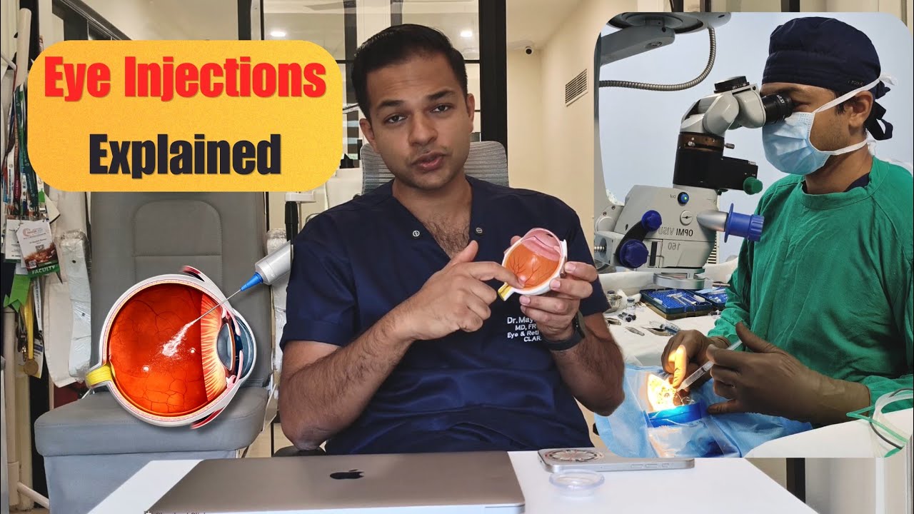
Medications injected into the eye to treat AMD, diabetic retinopathy, and retinal vein occlusion.
RETINA SURGERY FAQs